The art of illustrating what's inside our bodies
ALBANY, New York — Bill Westwood's work as a medical illustrator sits somewhere between art and science. The Albany, New York, illustrator has spent more than 50 years depicting different parts and processes inside the body for doctors, advertising agencies and pharmaceutical companies.
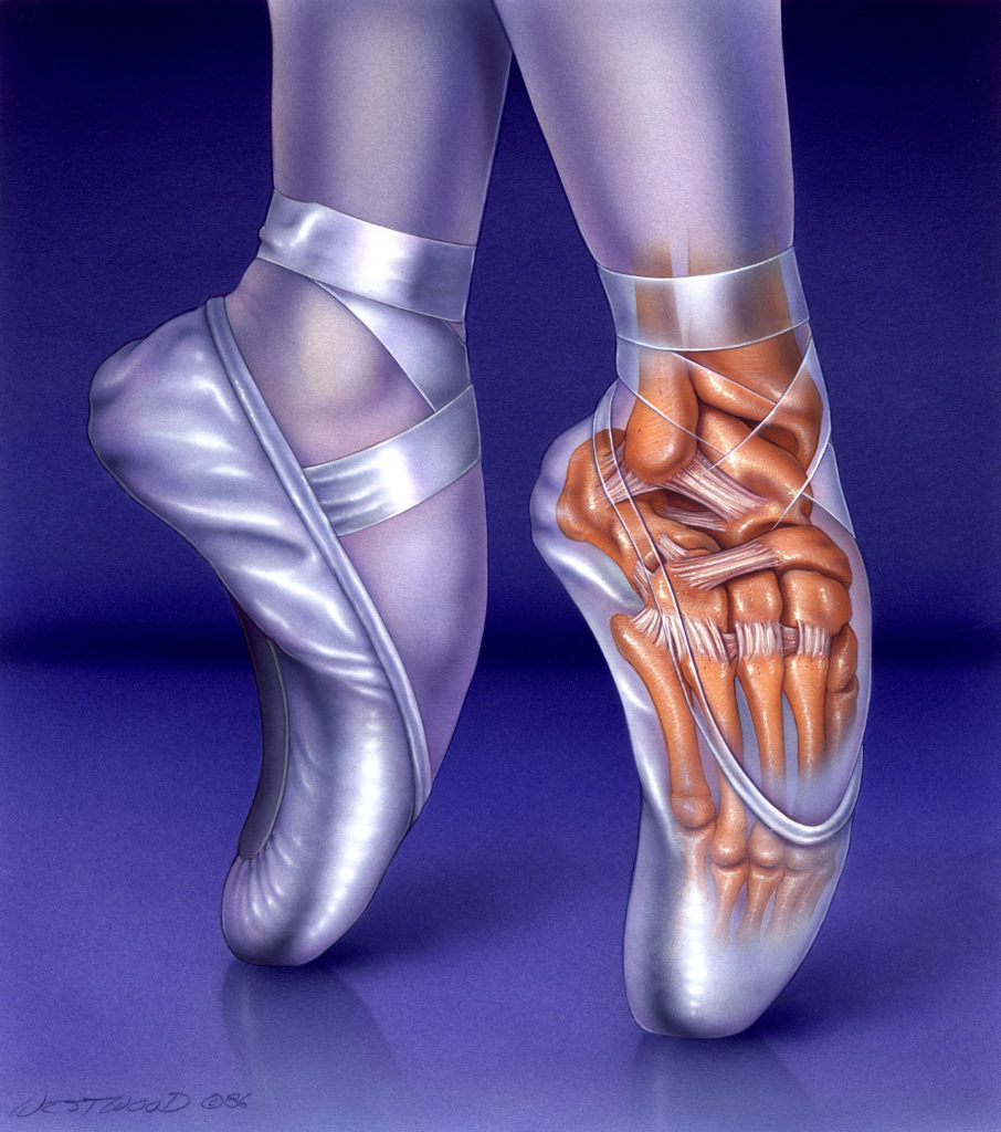
There's his instructional illustration of a surgeon removing a stomach in a gastrectomy. With editorial flair, Westwood pictures how the foot bones are compressed inside a ballerina's pointe shoe.
"I've gotten interested in the whole idea [of] being able to create accurate, educational and persuasive images that teach people about some of these injuries, that teach people about some of these surgeries," he said.
Westwood, who currently works out of a studio in Albany, said the work is different each day, including rendering images of injuries in legal cases. Westwood has been drawing medical illustrations since 1967 — and he has a design teacher from long ago to thank.
Decades earlier, Westwood was flabbergasted when he received a "C" in an undergraduate design class, so he decided to confront his teacher about the low grade. Little did Westwood know that the resulting conversation would introduce him to the field of medical illustration.
"That one design teacher changed my life," he said. Westwood took the advice of the professor and enrolled in a Georgia school that specialized in the work.
The school also only accepted four students per year; those students had to excel in both art and science. During his last two years at a liberal arts college in the same state, he "had never worked so hard in my life" to get in — and he was eventually accepted to the school.
Westwood showed WMHT how research-heavy the job can be. When he's ready to create a medical illustration, he starts by focusing on a sketch of the work. Once that gets laid out, he puts his sketch in a scanner and opens it on his computer. From there, he'll fill in the details, like airbrushing a red blood cell.
Westwood said his job is extremely rewarding and, even after 52 years, he has no plans to retire.
This report originally appeared on WMHT's "AHA! A House for Arts."
Support Canvas
Sustain our coverage of culture, arts and literature.


As the rainbow array outside Boston’s Institute of Contemporary Art suggests, artist Derrick Adams is accentuating the positive. He celebrates…

One of the biggest hits on Broadway right now is a reimagined version of “Cats,” the legendary musical about a…

Former President Barack Obama, joined by three former presidents, celebrated the opening of his presidential museum in Chicago in an…

A century ago, Black physicians built hospitals, clinics and medical schools across the South – only to see them dismantled…

The Shockoe Institute in Richmond, Virginia, opened its doors this spring to try to open minds about the enduring impact…

The United States is preparing to mark 250 years since its founding, a milestone often framed as a celebration of…

Globally celebrated South African jazz icon Abdullah Ibrahim, who performed at Nelson Mandela ’s 1994 presidential inauguration, has died at…

Banks contends in the lawsuit filed Saturday that they edited her interviews to create a false narrative.

British artist and painter David Hockney, one of the most celebrated art icons of the 20th and 21st centuries, died…

The Bruce Springsteen Center for American Music at Monmouth University features exhibits dedicated to one of New Jersey’s most famous…

Taylor Swift became the youngest woman ever inducted into the Songwriters Hall of Fame Thursday night at age 36.

A judge on Friday denied a request from the Kennedy Center to pause a ruling ordering President Donald Trump’s name…

Devlin Barrett has covered federal law enforcement for more than two decades. His new book pulls back the curtain on…

Should it reflect the host countries? Be a global banger? Value a chant-along chorus? There's an argument to make for…

Leo called Gaudí’s unfinished temple, one of the world’s most visited monuments, a “sign of unity and harmony for all…